

How to Interpret a Normal Pediatric Chest X-Ray: : What Every Parent Should Know
For many parents, hearing that their child needs an X-ray can spark worry. Even if the scan is routine, it’s natural to feel anxious about what doctors might find or what the images might show. Understanding the basics of pediatric chest radiology—and what a normal pediatric chest X-ray in radiology typically looks like—can help bring clarity and reassurance during medical visits.
This guide walks you through the fundamentals of how radiologists interpret a normal pediatric chest X-ray, what key structures they evaluate, how pediatric images differ from adult ones, and what normal childhood variations you may see in an image. In addition, you will learn how pediatric-focused teleradiology specialists support families and healthcare providers with accurate, timely readings.
📊
Pediatric Chest Imaging – A Primer
PowerPoint Presentation (.pptx)
The Importance of Getting a Pediatric Chest X-Ray
A chest X-ray is one of the most common imaging studies performed in children. It helps clinicians evaluate symptoms such as cough, fever, chest pain, breathing difficulties, or concerns about congenital heart or lung conditions. Unlike CT scans, chest X-rays use very low radiation doses and offer a quick, painless way to visualize internal structures.
In pediatric chest radiology, the goal is not only to detect abnormalities but also to reassure caregivers when an image is normal. Understanding what radiologists look for can help parents feel informed and empowered.
Evaluating Lung Expansion and Lung Fields
Lung appearance is one of the first features radiologists assess on a normal pediatric chest X-ray in radiology.

Lung Expansion
Radiologists check how fully the lungs inflate. In a normal pediatric chest X-ray:
- Both lungs should appear expanded and symmetrical.
- The diaphragm—a dome-shaped muscle under the lungs—should sit at a consistent height on both sides.
- Lung margins should extend downward toward the diaphragm without appearing overly flattened or excessively elevated.
Lung Fields
The “lung fields” refer to the areas visible on the film where air appears black. In a normal study:
- There should be uniform darkness across both lungs.
- No hazy patches, bright spots, or streaks should interrupt the lung fields.
- Blood vessels should fan out evenly from the center of the chest toward the outer edges.
Children often have more visible blood vessels than adults, which is entirely normal and reflects their developing circulatory system.
Heart Size and Shape
Another key component of pediatric chest radiology is evaluating the heart.
Heart Size
A child’s heart normally appears relatively larger in proportion to the chest compared to an adult’s. Radiologists measure the “cardiothoracic ratio”—the width of the heart compared to the width of the chest:
- In infants, the heart may appear up to 60% of the chest width.
- By school age, this ratio becomes closer to adult norms (around 50%).
A slightly larger-appearing heart in young children is not an automatic sign of disease.
Heart Shape
Radiologists also look for:
- Smooth heart borders
- Normal appearance of the heart chambers
- Clear visualization of the aortic arch and pulmonary vessels
The overall silhouette should appear well-defined without bulging or unusual contour changes.
Airway Structures and the Mediastinum
The airway and the mediastinum (the central area of the chest) tell radiologists a great deal about a child’s respiratory health.

Trachea and Bronchi
On a normal pediatric chest X-ray:
- The trachea should appear midline (centered).
- The main bronchi should branch symmetrically.
- No narrowing, bending, or blockage should be visible.
Because children have softer cartilage, the trachea may appear slightly more flexible or tapered—another normal pediatric finding.
Mediastinum
This area contains the heart, major blood vessels, thymus gland, and other structures. In children, the thymus gland is often prominent and can appear as:
- A soft, triangular or sail-shaped shadow
- Often mistaken for a mass by parents unfamiliar with pediatric imaging
But this “large-looking” thymus is a classic normal feature of pediatric chest radiology.
Bone Development and Skeletal Findings
The ribs, clavicles, spine, and shoulder joints provide valuable context.

Normal Bone Features
Radiologists look for:
- Normal alignment of the spine and ribs
- Even spacing between ribs
- A smooth clavicle outline
- No fractures or unusual curvatures
Children’s bones contain growth plates—lines or gaps near the ends of bones. These are normal and indicate healthy skeletal development.
Common Normal Variations
Some features that may appear concerning to parents but are common in normal pediatric chest X-rays include:
- Visible growth plates
- Slight forward curvature of the spine (kyphosis)
- Uneven rib visibility if the child moved or took a shallow breath
- Mild rotation if the child was wiggly during imaging
Movement is very common in young children and can influence how certain structures appear, which is why pediatric radiologists use specialized techniques to interpret images accurately.
How Pediatric Imaging Differs from Adult Imaging
When reading a normal pediatric chest X-ray in radiology, specialists use different reference points compared to adult X-rays because children are still developing.
Key Differences
- Proportionally larger thymus: Prominent in infants and toddlers; nearly invisible in adults.
- Higher heart-to-chest ratio: A normal developmental feature.
- More visible pulmonary markings: Reflects normal lung growth and blood flow.
- Rapid developmental changes: A normal X-ray for a 3-month-old looks different from a normal X-ray for a 10-year-old.
- Bone flexibility and growth plates: Present only in children.
- Increased movement artifacts: Children often cannot hold still or hold deep breaths.
Because of these differences, pediatric chest radiology requires dedicated expertise to distinguish normal variations from concerning findings.
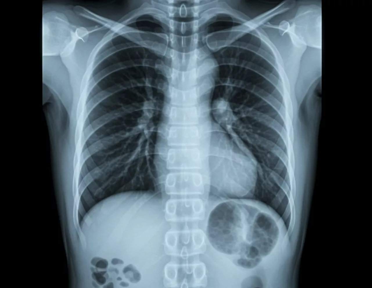
Putting It All Together: What a Normal Pediatric Chest X-Ray Should Show

Parents reviewing an X-ray image may notice:
- A large-looking heart
- A cloud-like structure near the upper chest (the thymus)
- Extra or prominent lung markings
- Slightly tilted or rotated images
These are often completely normal and expected in pediatric imaging. Radiologists trained in child imaging understand these variations and interpret them with great care.
A normal pediatric chest X-ray in radiology generally reveals:
- Clear, well-expanded lungs
- A heart size appropriate for the child’s age
- Airway structures that are open and midline
- Bones with visible growth plates and normal development
- Normal pediatric variations such as a prominent thymus
These features indicate healthy respiratory and cardiac systems and support your child’s medical team in ruling out concerns safely and quickly.
Specialty Focused Radiology: Your Partner in Pediatric Teleradiology
Interpreting pediatric imaging requires skill, experience, and a deep understanding of developmental anatomy. Specialty Focused Radiology in South Florida is proud to serve as a trusted partner in pediatric chest radiology for hospitals, clinics, and pediatric practices nationwide. Our board-certified pediatric radiologists provide expert teleradiology services such as remote readings, diagnostic interpretations, and comprehensive reports designed to support timely and accurate treatment decisions. We combine advanced imaging technology with compassionate, family-centered care—ensuring every child receives the highest level of diagnostic expertise no matter where they live.
Contact Us for Expert Pediatric Imaging Support
If you need assistance interpreting a pediatric X-ray or want to learn more about how our teleradiology services can support your child’s healthcare team, Specialty Focused Radiology is here to help. Contact us today to schedule an appointment, request a consultation, or speak with a pediatric imaging specialist who can guide you through the process with clarity, accuracy, and care.
FAQs
How is a pediatric chest X-ray different from an adult chest X-ray?
Pediatric X-rays show different proportions, including a larger thymus, a relatively larger heart, and more visible lung markings. Radiologists who specialize in pediatric chest radiology use these age-specific differences to distinguish normal findings from potential concerns. How much radiation does a pediatric chest X-ray use?
Pediatric chest X-rays use very low doses of radiation, often comparable to what a child experiences naturally over a few days of normal environmental exposure. Modern equipment and pediatric protocols ensure imaging is as safe as possible. What do radiologists look for in a normal pediatric chest X-ray?
They examine lung expansion, airflow, heart shape and size, airway alignment, bone growth, and the appearance of the thymus. Each structure is evaluated using pediatric-specific norms to ensure the image reflects healthy development. When should my child have a chest X-ray?
A chest X-ray is typically recommended when a child has persistent cough, fever, breathing difficulty, chest pain, or concerns about congenital heart or lung issues. Your pediatrician will determine if imaging is appropriate based on symptoms and exam findings.
Pediatric X-rays show different proportions, including a larger thymus, a relatively larger heart, and more visible lung markings. Radiologists who specialize in pediatric chest radiology use these age-specific differences to distinguish normal findings from potential concerns.
Pediatric chest X-rays use very low doses of radiation, often comparable to what a child experiences naturally over a few days of normal environmental exposure. Modern equipment and pediatric protocols ensure imaging is as safe as possible.
They examine lung expansion, airflow, heart shape and size, airway alignment, bone growth, and the appearance of the thymus. Each structure is evaluated using pediatric-specific norms to ensure the image reflects healthy development.
A chest X-ray is typically recommended when a child has persistent cough, fever, breathing difficulty, chest pain, or concerns about congenital heart or lung issues. Your pediatrician will determine if imaging is appropriate based on symptoms and exam findings.