
Radiology plays a central role in modern medicine, offering clinicians a spectrum of imaging tools to visualize anatomy, assess pathology, and guide decision-making. These radiology modalities—from basic X-ray to advanced MRI, PET, and subspecialty techniques—each offer unique advantages. Understanding the differences among them is essential for clinicians, technologists, and referring providers who must choose the most appropriate test for accurate, efficient diagnosis.
This guide provides a clear overview of the different modalities in radiology, explaining how they work, their strengths and limitations, and which clinical scenarios benefit most from each. With the right modality, diagnostic confidence increases significantly—leading to better patient care, fewer repeat exams, and more focused treatment planning.
Foundational Radiology Modalities
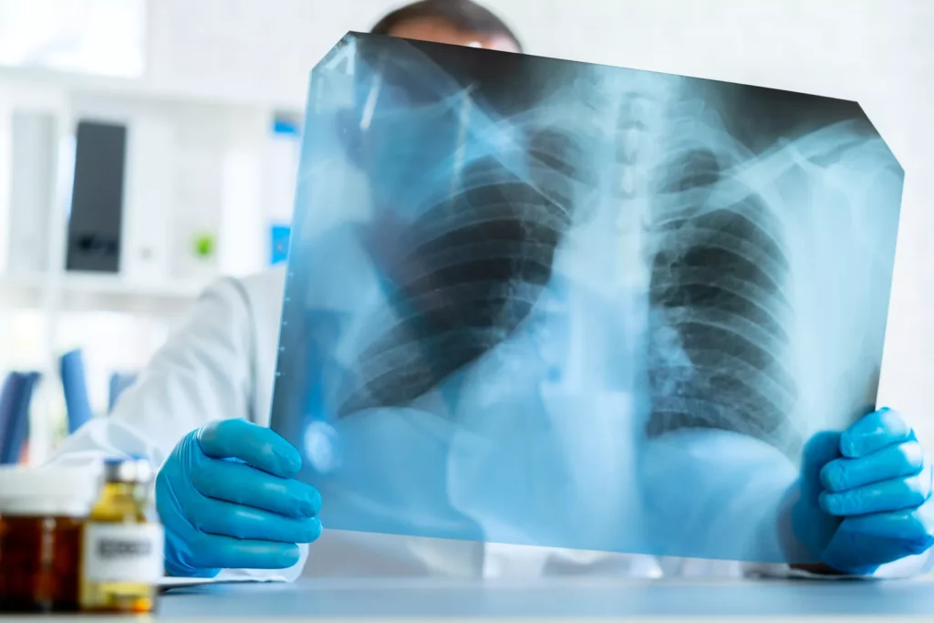
X-Ray (Radiography)
How it works:
X-ray uses ionizing radiation to create 2D images of internal structures based on tissue density.
Ideal use cases:
- Bone fractures
- Chest evaluation (pneumonia, effusions)
- Joint alignment
- Abdominal obstruction screening
Strengths:
- Fast, widely available, low cost
- First-line tool for many acute conditions
Limitations:
- Limited soft-tissue detail
- Overlapping anatomy can obscure pathology
Diagnostic role:
X-ray often provides the first clue that guides further imaging selection, forming the foundation for most imaging pathways.
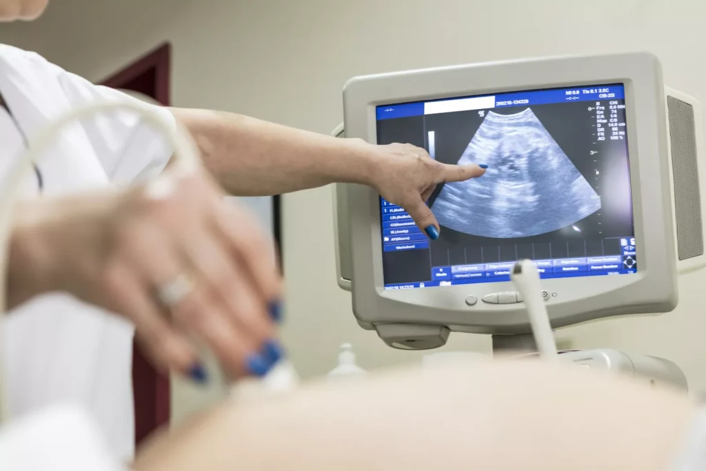
Ultrasound
How it works:
Ultrasound uses high-frequency sound waves to generate real-time images. No radiation is involved.
Ideal use cases:
- Obstetrics
- Gallbladder disease
- Thyroid and soft-tissue evaluation
- Vascular studies (DVT, arterial stenosis)
Strengths:
- Dynamic, radiation-free, portable
- Excellent for superficial structures and fluid collections
Limitations:
- Operator-dependent
- Limited acoustic penetration in obese patients or gas-filled areas
Diagnostic role:
Ultrasound is a first-line modality for abdominal, pelvic, and vascular concerns and is especially valuable for guiding procedures.
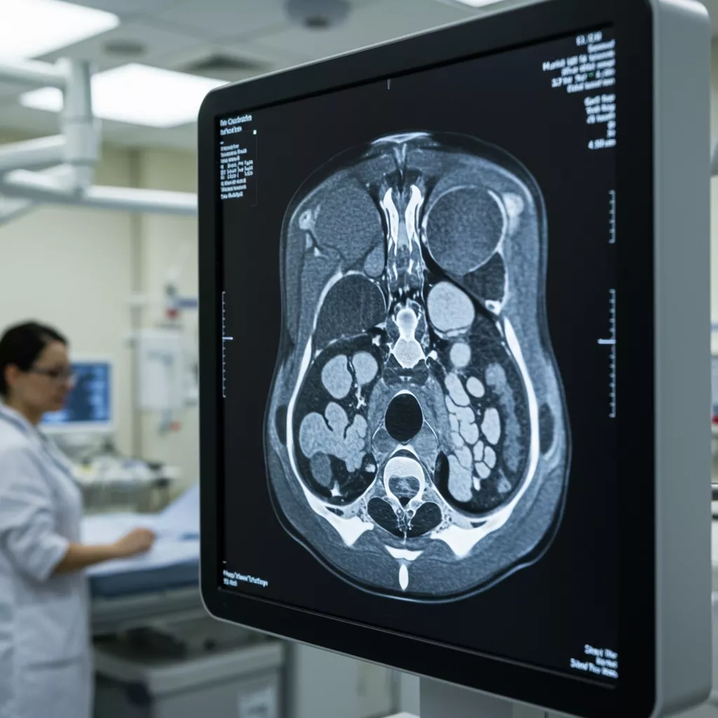
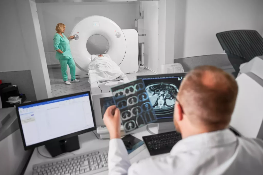
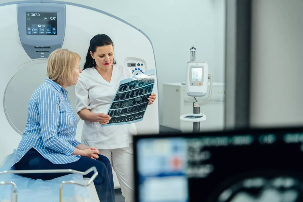
Computed Tomography (CT)
How it works:
CT uses rotating X-rays to produce cross-sectional images, offering excellent spatial detail.
Ideal use cases:
- Trauma imaging
- Stroke evaluation
- Pulmonary embolism
- Abdominal and pelvic pain
- Oncology staging
Strengths:
- Rapid acquisition
- High-resolution detail of bone, organs, and vasculature
- Excellent for acute pathology
Limitations:
- Uses ionizing radiation
- Iodinated contrast may not be suitable for all patients
Diagnostic role:
CT provides definitive answers in many urgent scenarios and offers precise details that surpass standard radiography.
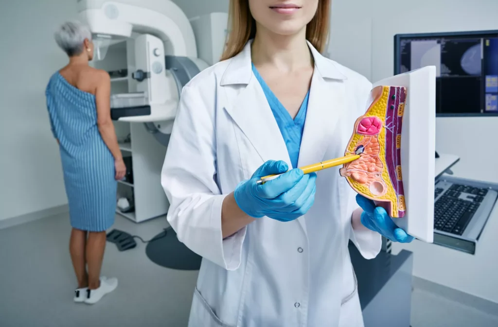
Mammography
How it works:
Mammography is a specialized radiology modality that uses low-dose X-ray technology to image breast tissue. It is designed to detect subtle differences between normal and abnormal structures, making it essential for early cancer detection.
Ideal use cases:
- Breast cancer screening
- Diagnostic evaluation of breast lumps, pain, or nipple changes
- Assessing calcifications, masses, or architectural distortion
Strengths:
- Proven effectiveness in detecting early-stage breast cancer
- Low radiation dose
- Highly standardized across facilities
Limitations:
- Reduced sensitivity in dense breast tissue
- May require additional imaging such as ultrasound or MRI for clarification
Diagnostic role:
As the cornerstone of breast imaging, mammography enables earlier detection of malignancy, allowing improved outcomes and more precise diagnostic pathways when combined with breast ultrasound or MRI.
Advanced Radiology Modalities
Magnetic Resonance Imaging (MRI)
How it works:
MRI uses magnetic fields and radiofrequency waves to create detailed images, particularly of soft tissues.
Ideal use cases:
- Brain and spine
- Musculoskeletal injuries
- Tumor characterization
- Cardiac and vascular assessment
Strengths:
- Superior soft-tissue contrast
- No ionizing radiation
- Multiparametric capabilities
Limitations:
- Longer exam times
- Contraindications for some implants
- Higher cost and less availability than CT
Diagnostic role:
MRI often provides the decisive detail needed to confirm or exclude complex pathologies, especially in neurology and orthopedics.
Positron Emission Tomography (PET)

How it works:
PET tracks radiotracer uptake, often combined with CT or MRI to correlate metabolism with anatomy.
Ideal use cases:
- Oncology staging
- Cardiac viability
- Neurologic disorders (e.g., dementia evaluation)
Strengths:
- Highly sensitive for malignancy and inflammation
- Critical for treatment planning and monitoring
Limitations:
- Expensive and less widely available
- Requires specialized tracers
Diagnostic role:
PET is indispensable for cancer management and functional brain imaging.
Fluoroscopy
How it works:
Fluoroscopy uses continuous X-ray to capture real-time motion.
Ideal use cases:
- GI tract studies (swallow, UGI, barium enema)
- Interventional procedures
- Joint injections (arthrography)
Strengths:
- Dynamic assessment
- Valuable procedural guidance
Limitations:
- Continuous radiation exposure
- Limited soft-tissue detail
Diagnostic role:
Fluoroscopy is primarily procedural or functional, offering insights no static image can provide.
How Modality Selection Improves Diagnostic Accuracy
Choosing the right imaging modality is essential for achieving clear, reliable diagnostic results. Each modality offers unique strengths—such as CT for rapid evaluation, MRI for superior soft-tissue detail, ultrasound for dynamic assessment, and mammography for early breast cancer detection—making proper selection critical for answering specific clinical questions.
When clinicians align clinical suspicion with the most suitable modality, they reduce uncertainty, avoid unnecessary tests, and obtain more actionable information on the first attempt, leading to faster and more confident decision-making.
Subspecialty Considerations: Tailoring Modalities to Clinical Focus
Subspecialists select modalities based on age, anatomy, urgency, and diagnostic complexity, ensuring tailored, high-value imaging.
Musculoskeletal Imaging (MSK)
- X-ray: First-line for trauma and degenerative disease
- MRI: Gold standard for soft-tissue injuries and occult fractures
- Ultrasound: Excellent for tendon pathology and dynamic assessment
Neuroimaging
- CT: Acute stroke, hemorrhage, trauma
- MRI: Tumors, demyelination, epilepsy, chronic ischemia
Breast Imaging
- Mammography: Screening and diagnostic evaluation
- Ultrasound: Characterizing masses and guiding biopsies
- Breast MRI: High-risk screening, implant evaluation
Cardiothoracic Imaging
- CT/CTA: Coronary arteries, pulmonary embolism, interstitial lung disease
- MRI: Cardiac function, viability, congenital heart disease
Pediatric Radiology
- Ultrasound: Preferred for appendicitis, hips, and abdomen
- MRI: Excellent alternative to CT to reduce radiation
- X-ray: Limited but essential for trauma and respiratory evaluation
Major Radiology Modalities Compared
| Modality | Radiation? | Best For | Key Strength | Primary Limitation |
| X-ray | Yes | Bones, chest | Fast, inexpensive | Limited detail |
| Ultrasound | No | Soft tissues, OB, vascular | Dynamic, portable | Operator-dependent |
| CT | Yes | Acute conditions, trauma | High resolution, rapid | Radiation + contrast |
| MRI | No | Neuro, MSK, soft tissues | Superior detail | Cost, time |
| Nuclear Medicine | Yes | Functional imaging | Early disease detection | Low spatial resolution |
| PET | Yes | Oncology, neurology | Sensitive for malignancy | Limited availability |
| Fluoroscopy | Yes | GI studies, procedures | Real-time imaging | Continuous radiation |
Specialty Focused Radiology: Your Partner in Precision and Diagnostic Confidence
Here at Specialty Focused Radiology, we deliver high-quality, subspecialty-driven teleradiology interpretations designed to strengthen diagnostic accuracy across all major radiology modalities. Our team includes experienced MSK, neuro, body, cardiothoracic, and emergency radiologists who provide precise, clinically meaningful reports for X-ray, CT, MRI, ultrasound, and advanced subspecialty imaging. We emphasize clarity, consistency, and rapid turnaround—ensuring referring clinicians receive the exact level of detail and expertise needed to make confident decisions.
By pairing deep subspecialty knowledge with dependable teleradiology support, we help healthcare organizations elevate their imaging quality while maintaining efficiency. Whether interpreting routine studies or managing complex diagnostic questions, Specialty Focused Radiology serves as a reliable extension of your clinical team, improving outcomes through expert, modality-specific insight.
Enhance Your Diagnostic Confidence. Contact Specialty Focused Radiology Today
Understanding the different modalities in radiology is essential for making accurate, timely, and confident clinical decisions. With the increasing complexity of modern imaging, having expert support is more important than ever. At Specialty Focused Radiology in South Florida, we provide the subspecialty expertise clinicians rely on to interpret imaging accurately across all major radiology modalities.
If your practice is seeking clearer answers, dependable support, and high-quality teleradiology interpretations, contact Specialty Focused Radiology today to learn how our team can enhance your diagnostic confidence.
FAQs
What are radiology modalities?
Radiology modalities are the different imaging techniques used to visualize structures inside the body, such as X-ray, ultrasound, CT, MRI, and nuclear medicine. Each modality uses unique technology to evaluate anatomy or physiology. Understanding how they differ helps clinicians choose the most accurate test for each clinical scenario.
How do I know which radiology modality is best for my patient?
The best modality depends on the symptoms, suspected diagnosis, and the level of detail required. Clinicians typically follow established imaging pathways or guidelines to match concerns with the appropriate test. Consulting with radiologists can further improve modality selection and diagnostic accuracy.
How can teleradiology benefit my practice or imaging center?
Teleradiology ensures your patients receive timely interpretations, even during high-volume or after-hours periods. It provides access to subspecialty expertise that may not be available onsite. This supports better workflow, reduced delays, and more consistent diagnostic quality.
What is the best imaging modality for cancer detection?
The best modality depends on the type of cancer. CT and MRI provide detailed anatomical information, while PET excels at detecting metabolic activity associated with malignancy. Often, a combination of modalities offers the most accurate assessment.