
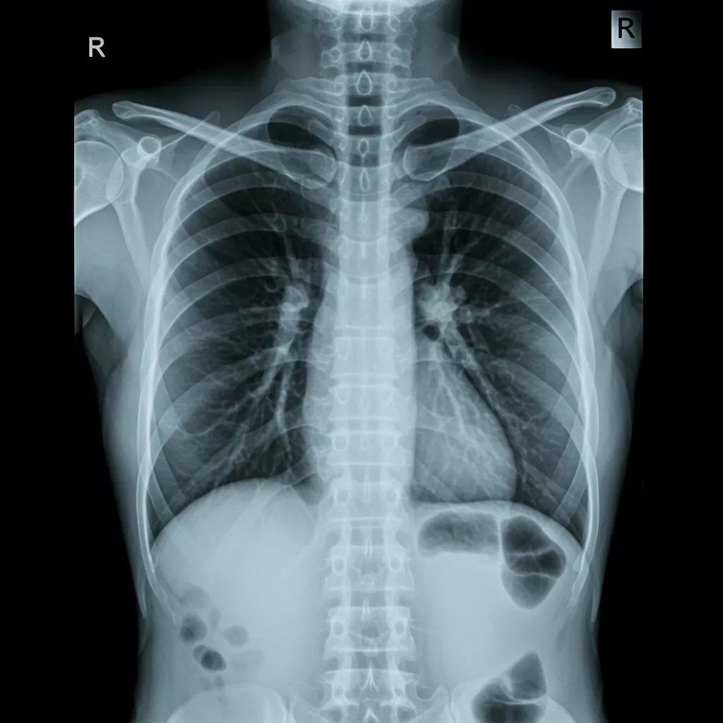
A chest X-ray (CXR) is one of the most widely used diagnostic imaging tools in medicine. It offers a fast, noninvasive look at the heart, lungs, airways, bones, and surrounding soft tissues. Whether used in emergency settings, routine evaluations, or ongoing monitoring, understanding the difference between a normal chest X-ray and an abnormal chest X-ray is essential for guiding accurate clinical decisions. This comparison guide breaks down key features to help readers better understand chest X-ray normal vs abnormal findings and what clinicians look for during interpretation.
What a Chest X-Ray Evaluates
A chest X-ray captures vital structures within the thoracic cavity, providing visual information about:
- Lungs: Air-filled spaces, vascular markings, and signs of infection or fluid
- Heart and Mediastinum: Size, shape, and potential widening or displacement
- Pleural Spaces: Indicators of fluid (effusion), air (pneumothorax), or thickening
- Bones and Chest Wall: Rib fractures, spinal alignment, or deformities
- Diaphragm: Position, contour, and movement abnormalities
- Medical Devices: Placement of lines, tubes, or implants
Because so many critical structures are evaluated, differentiating normal vs abnormal chest X-ray findings is crucial for early detection, appropriate treatment, and patient safety.
Normal Chest X-Ray: Key Features
A normal chest X-ray includes several hallmark characteristics demonstrating healthy thoracic anatomy. Some of the most important features include:
1. Clear, Symmetric Lung Fields
- Lungs appear radiolucent (dark) with normal vascular markings.
- No areas of consolidation, mass effect, or interstitial thickening.
2. Normal Heart Size and Contours
- Cardiothoracic ratio is within normal limits (typically <50% on PA view).
- Heart borders are sharp and well-defined.
3. Proper Mediastinal Width
- Trachea is midline.
- Mediastinum appears appropriately sized without widening or displacement.
4. No Pleural Abnormalities
- No visible fluid, air, or pleural thickening.
- Costophrenic angles are sharp and unobstructed.
5. Normal Bones and Soft Tissues
- Ribs, clavicles, and spine show no fractures or lesions.
- Soft tissues appear symmetric without masses or subcutaneous emphysema.
Recognizing these baseline findings provides the foundation for detecting abnormalities quickly and accurately.
Abnormal Chest X-Ray: Common Findings to Watch For

An abnormal chest X-ray can present a wide range of patterns, each suggesting different potential diagnoses. Understanding these patterns helps clinicians compare normal vs abnormal chest X-ray features with confidence.
1. Consolidation or Airspace Opacity
- Appears as white or opaque areas within the lung.
- Common causes: pneumonia, pulmonary hemorrhage, aspiration.
2. Interstitial Abnormalities
- Reticular or nodular patterns suggest interstitial lung disease, pulmonary edema, or viral infections.
3. Cardiomegaly or Abnormal Heart Contours
- Enlarged cardiac silhouette may indicate heart failure, pericardial effusion, or cardiomyopathy.
4. Pleural Effusion or Pneumothorax
- Effusion: blunted or obscured costophrenic angles with fluid layering.
- Pneumothorax: hyperlucent (very dark) area lacking lung markings, often with visible pleural line.
5. Masses, Nodules, or Calcifications
- Focal lesions may represent tumors, granulomas, or metastases.
6. Abnormal Device Positioning
- Malpositioned central lines, tubes, or pacemaker leads can cause complications requiring immediate attention.
By comparing these findings to the expected anatomy of a normal chest X-ray, clinicians can more easily distinguish clinically significant changes.
Chest X-Ray Normal vs Abnormal: A Systematic Approach
To accurately assess chest X-ray normal vs abnormal images, radiologists and clinicians often follow a structured review method:
- Airway – alignment and patency
- Bones – fractures, lesions, symmetry
- Cardiac – size, borders, contours
- Diaphragm – shape, height, clarity
- Effusions/Extrathoracic tissue – pleural spaces and soft tissues
- Fields of the lungs – opacities, vascularity, infiltrates
- Gadgets – checking placement of lines/devices
This systematic approach reduces the chances of missing subtle abnormalities and improves diagnostic reliability.
Expert Chest X-Ray Interpretations from Specialty Focused Radiology
At Specialty Focused Radiology in South Florida, our team of highly experienced teleradiologists specializes in providing accurate, reliable readings and interpretations across normal vs abnormal chest X-ray studies. We routinely handle a full spectrum of cases—from routine outpatient exams to urgent findings and complex thoracic conditions. Our teleradiologists excel at identifying subtle abnormalities, delivering clear and timely reports, and supporting clinicians with actionable diagnostic guidance. With deep expertise in thoracic imaging, we ensure precision and consistency in every chest X-ray interpretation.
Contact Specialty Focused Radiology for Expert Chest X-Ray Interpretations
Here at Specialty Focused Radiology, we are committed to delivering that precision—providing dependable, expert interpretations that elevate patient care and strengthen your diagnostic workflow. If your practice is ready for faster, more accurate, and highly reliable chest X-ray readings, contact us here at Specialty Focused Radiology today and experience the difference true subspecialty expertise makes.
FAQs
What does a normal chest X-ray look like?
A normal chest X-ray shows clear lung fields, a heart of normal size, and well-defined ribs and diaphragm. There are no unusual shadows, masses, fluid buildup, or signs of infection or injury.
What does an abnormal chest X-ray look like?
An abnormal chest X-ray shows findings that differ from expected anatomy, such as white patches, dark areas, enlarged organs, or irregular shapes. These abnormalities may indicate infections, lung disease, heart conditions, or structural problems.
Does an abnormal chest X-ray always mean serious disease?
No, an abnormal result does not automatically indicate a serious condition. Many findings require correlation with symptoms, blood tests, or additional imaging before a diagnosis is made.
What should I do if my chest X-ray is abnormal?
You should follow up with your healthcare provider for interpretation and next steps. Additional tests or treatments may be recommended based on the findings and symptoms.